
Advanced Technology
Digital X-Rays:
Digital X-rays offer greater precision because the images can be viewed on a computer monitor, allowing clinicians to enhance, magnify, and adjust contrast or brightness for better diagnostic accuracy.
Kavo(3D Diagnostic Imaging)
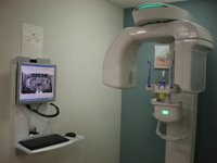 Kavo is the cone beam 3D + panoramic imaging solution for general dentists and specialists who want to place and restore implants, perform surgical procedures, and simply provide better care with more confidence and efficiency and greater patient engagement than before. With this exclusive treatment planning solution, Kavo provides powerful yet easy to use tools placing and restoring implants, including abutments and crowns, performing implant surgery, complex extractions, treating endodontic (root canal) cases and better visualizing pathology. Rich visual images come to life and engage patients in their treatment to assist in better understanding and acceptance of proposed plans. A powerful diagnostic and treatment tool it allows complex procedures to be done quickly, and with greater confidence and accuracy. 3D CT Diagnostic Imaging clearly facilitates better treatment planning in dental care for many situations.
Our 3D cone beam dental scanner is approved by the FDA and is often referred to as a “dental CT (conebeam) scanner”. Dental cone beam scanners use much less radiation as opposed to medical CT scanners and more accurate, diagnostic images for implant placement, removal of impacted wisdom teeth and more.
Cadent iTero Digital Impression System
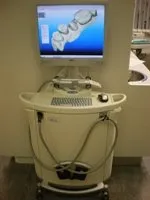 An accurate impression is fundamental to a successful outcome in restorative dentistry. Conducting digital impression procedures with Cadent iTero provides consistent, superior results, as well as numerous other benefits for the clinician, patient, and dental laboratory.
The iTero user interface guides users through every step of the scanning process. The time to complete the scanning sequence is routinely just 3-5 minutes, about the time required for conventional impression materials to set. And, with no time devoted to preparing impressions trays or clean-up, the time savings are predictable. Better still, the unmatched precision of iTero digital impressions provides crowns that require minimal adjustment, another source of predictable time savings.
Data files of the digital impression are sent electronically, enhancing the speed of communication involved in restoration fabrication. Furthermore, should questions arise, a digital file is available for the laboratory and dentist to reference while consulting about a case online.
Dental practices using the iTero digital impression system will enjoy a distinct advantage over those adhering to the conventional impression method. With iTero, patients become engaged in the process as they see their dental work on-screen. This provides the clinician with a wonderful educational tool that helps improve patient acceptance.
Laser Dentistry
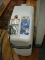 For patients who do not look forward to needles, drilling, or numbness, Laser Dentistry may be the right choice. Laser dentistry is one of dentistry’s latest advances. The laser delivers energy in the form of light. Depending on the intended result, this energy travels at different wavelengths and is absorbed by a “target.” In dentistry, these targets can be enamel, decay, gum tissue, or whitening enhancers. Each one absorbs a different wavelength of light while reflecting others. Laser dentistry can be used for both tooth and soft tissue related procedures. Oftentimes no local anesthesia is required. Unlike with the dental drill, with laser dentistry there is no heat or vibration, making the procedure quite comfortable for most patients. Healing is often much faster. Lasers can be used to diagnose cavities. They can find hidden decay in teeth in early stages, and in some cases the decay can be reversed through hygiene and fluoride treatment and may never need filling.
Areas of dental care that benefit from laser technology:
- Cavity diagnosis and removal
- Periodontal, or gum related, care
- Pediatric procedures
- Apthous Ulcer treatment (canker sore)
- Frenectomy (tongue-tie release) without anesthesia or sutures that is very beneficial in the new-born to facilitate better nursing
- Root canals and apicoectomies
- Crown lengthening, gingivectomy and other gum corrections
Dental lasers have been shown to be safe and effective for treating both children and adults.